If you want to become a personal trainer, its a good to know that muscles will grow, or hypertrophy, when thoughtfully trained and when proper nutrients are supplied. But, exactly what biological system is responsible for this growth stimulus? Is it one or is it several of them? Is growth caused by the forces applied to the muscles through weight training or on a more cerebral level? Is resistance exercise merely a means by which we expose, through recruitment, as many muscle fibers as possible to some other stimulus responsible for growth? Let’s break it down.
Muscle Synthesis
All tissues undergo a formation period (synthesis) and a breakdown period (degradation), also known as turnover. This typically occurs in a state of homeostasis where very little variation occurs. Old tissues are replaced by new ones which look and perform essentially the same as their predecessor. However, muscular hypertrophy requires an imbalance in this equation as the period following the degradation results in the laying down and retention of a greater amount of tissue than was present previously.
D. A. Jones, et al., theorizes that there may be three possible mechanisms responsible for muscular growth:
- hormonal stimuli
- metabolic stimuli
- mechanical factors
We know there are a number of powerful hormonal reactions to exercise, but it is unlikely that hormonal stimuli are primarily responsible for muscle growth. Because of their generalized effects and the fact that muscular growth is typically limited to only those muscles which are trained, it would appear that hormones only work with some other localized muscular change with a greater influence on muscular growth.
There are situations in which localized hormonal changes in fibroblasts (material that becomes connective tissue) produce growth factors that may have an effect on the muscles in their vicinity. It’s not uncommon to “go for the burn” during intense, heavy training. This kind of training is typically associated with high concentrations of metabolites which result from the reactions that take place during energy production for muscular contractions (note: This burning sensation has conventionally believed to be lactic acid accumulation, however, there are people who cannot produce lactic acid due to a metabolic disorder, and yet experience the “burn” anyway).
However, these metabolites are typically associated with failure of the energy systems (muscular fatigue) rather than force production. They may induce an increase in mitochondria and/or capillary growth, but this contributes very little to hypertrophy. For example, marathon runners have extensive mitochondrial and capillary proliferation, yet they have weaker and smaller lower extremity muscles relative to athletes who train for hypertrophy.
High Force = Hypertrophy?
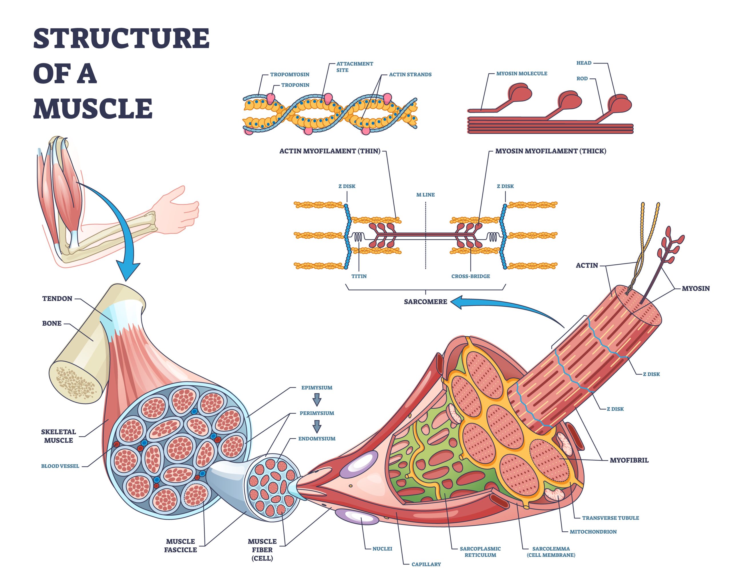
High-force mechanical stress, whether by contraction or repeated stretching, obviously plays a key role in the muscle growth equation. The role such stress plays varies across a few theories. Let’s revisit the structures and function of muscle tissue.
Myofibrils are the contractile components that allow for strength and performance of the muscles; they are composed of repeating sections of functional structures called sarcomeres that give skeletal muscle cells the appearance of striations. A myofibril is further composed of the protein myofilaments actin and myosin. These protein filaments possess the cross-bridges, or actual contractile units, along their length. You might visualize the myofilaments of actin and myosin as thin wire filaments that connect to each other, encased in a myofibril tube.
What makes muscle cells unique to other cells in the body is the fact that they have several nuclei and not just one, present along the entire length of the fiber and embedded in the cell membrane (sarcolemma).
It may cause the fracturing of myofilaments which split and regenerate to full size part of which is supported by electron micrograph photos.
The ribosomes within each muscle cell allow mRNA material (released from the DNA in the nucleus) and convert it to protein. Protein synthesis takes place by connecting one amino acid at a time so that it can be used as part of a larger protein.
In this fashion, the ribosomes become an “assembly line” that builds and repairs the contractile proteins, actin and myosin, in the damaged myofibrils, using available intracellular amino acids, resulting in the growth of tissue.
Heavy, intense, low-rep resistance training directly damages the actin and myosin in the myofibril. The “controlled” damage (or exercise) and repair (or recovery) of the actin and myosin is the key to optimizing myofibril growth. The process by which actin and myosin are repaired is commonly known as hypertrophic protein synthesis.
Althernatively, hypertrophy may also occur when heavy forces on the muscle tissue cause microtrauma within muscle cells, causing nearby satellite cells to divide and contribute their DNA material to the muscle cell nucleus. In this way, the damaged cell will synthesize more ribosomes and produce more protein for regeneration and hypertrophy. Hormonal growth factors are at play here as well; hepatocyte growth factor (HGF), a key regulator of satellite cell activity, has been shown to be the active factor in damaged muscle. It may be the impetus for satellite cells to travel to the damaged muscle area for rebuilding.
In any case, the purpose of the cell size growth is to prepare the cell for future challenges that are more likely to be met with a stronger, larger muscle fiber. The optimal approach for stimulating hypertrophy, or muscle cell size growth, is to apply heavy resistance forces within an exercise program.
What qualifies as “heavy”? How do we define a subjective term such as “heavy” which can be relative and also vary across organizations? According to the NFPT study and reference manual, hypertrophy will occur optimally in the 4-6 rep range utilizing loads at around 70-80% of 1RM for 1-6 sets.
This is a framework from which to start. Each body responds differently and qualified personal trainers will accurately assess and re-assess their clients to make sure any goals of hypertrophy are being achieved.
References
Jones, D.A., Rutherford, O.M., Parker, D.F. Physiological Changes in Skeletal Muscle as a Result of Strength Training. Quarterly Journal of Experimental Physiology. 74: 233-256. 1989.
https://pubmed.ncbi.nlm.nih.gov/24505026/
NFPT Study and Reference Manual: The Fundamentals for the Certified Personal Trainer, 7th Edition; pages 59-60 (2017).
https://www.unm.edu/~lkravitz/Article%20folder/musclesgrowLK.html#:~:text=Muscle%20growth%20occurs%20whenever%20the,controlled%20by%20complimentary%20cellular%20mechanisms.
As NFPT Blog Publisher from 2019 to 2024, and a veteran health and fitness trainer for over 20 years, Michele G Rogers, MA, NFPT-CPT, I-HTMAP, now serves as a Holistic Health Navigator and Practitioner, helping people improve their overall health utilizing Hair Tissue Mineral Analysis as a data-anchor. This functional medicine screening test enables clients to connect their physical symptoms to their emotional, cognitive, and spiritual health. She holds a master’s degree in applied health psychology from Northern Arizona University and multiple health certifications. Follow Michele on Instagram or find out more about her services at www.encompasspathways.com