The intrinsic muscles of the back control the vertabral column, i.e., the spine, and are situated deep below the extrinsic muscles which are responsible for movements of the thoracic cage and shoulder joints. The spinotransversales group are the first (top) layer of the deep intrinsic back muscles that contribute to cervical extension and rotation.
Every year, between 30 and 50% of the population suffers from disruptive neck pain. Worse, back pain is the single most disabling condition preventing sufferers from engaging in work or ADL’s, worldwide. An estimated 80% of the population will suffer from back pain at some point in their lives.
As personal trainers, it can be helpful to have a more detailed knowledge of the muscles that are often the source of chronic pain and dysfunction.
Understanding the Layers
All of the muscles of the back fall into one of three main groups:
- Superficial: The most superficial of extrinsic back muscles associated with shoulder motions, like the lats and rhomboids.
- Intermediate: The layer of extrinsic muscles beneath the superficial layer, which control the motions of the thoracic cage, i.e, the ribs: Includes serratus posterior inferior and superior.
- Deep: The deepest layer of intrinsic muscles associated with the vertebral column, head and neck.
Now, the deep, or intrinsic, group of back muscles is further broken down into those same exact category names as above. To maintain clarity, I will designate them with “intrinsic”:
- Superficial Intrinsic: The group called spinotransversales made up of the splenius capitis and splenius cervicis, both of which control the movements of the head and neck and extend from the upper spine and base of the skull, superficial to the deeper muscles of the neck. (the rest of this article discusses these two muscles)
- Intermediate Intrinsic: The group called erector spinae responsible for spinal flexion/lateral flexion.
- Deep Intrinsic: The group called transversospinales (yes this is different from the spinotransversales above!) comprised of short muscles running along the transverse and spinous processes of the vertebral column.
The Spinotransversales Group
The following two muscles form the spinotransversales and work in conjunction with the three other layers of cervical extensors (five other muscles total) and the four smaller craniocervical extensors which connect the base of the skull to the cervical spine in order to produce cervical flexion and rotation.
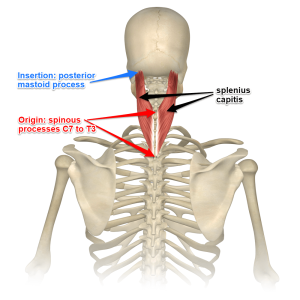
 Splenius Capitis
The word splenius comes from the Greek spléníon, meaning “bandage”. I supposes these two muscles might appear as Band-Aids at the back of the neck?
This transversospinales lie just beneath the trapezius muscle and levator scapulae (the most superficial and more global shoulder/cervical muscles) and are responsible for ipsilateral rotation and lateral flexion of the head (capitis!) as well as bilateral cervical extension. It attaches from the spinous processes of T3/T4 and C7, invests in the nuchal ligament, (fibrous attachment that runs atop the spine) and ascends to the mastoid process and occipital bone on the base of the skull.

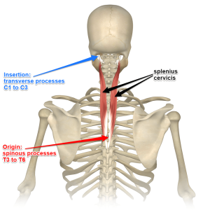
Splenius Cervicis
This muscle is longer than capitis and articulates along more of the cervical spine. This muscle also controls cervical extension, lateral flexion as well as rotation of the head. It attaches beneath the trapezius and rhomboids from the T6-T3 spinous process up to the transverse process of C3/4 and C1.
Training Considerations for Spinotransversales
Overactivity in this muscle group will present as forward head or “text neck” along with the other more superficial extensors. (interestingly, the deeper transversospinales tend to be underactive). Expect to see this more and more in our youth especially. Ignoring the cervical dysfunction when executing a fitness plan with a client will ultimately translate into pain, (or more likely more pain), and reinforcement of the faulty movement patterns as the stronger, more active muscles continue to take over for the weaker, inactive muscle groups.
In the case of the spinotransversales being short or overactive, it will be necessary to help the client release the muscles first (foam rolling and/or targeted release with a ball will work). Then lengthen it with a purposeful stretch by initiated the opposing actions that the muscles perform. In this case, tuck the chin activating the deep cervical flexors, pull the top of forward and slightly laterally to stretch one side and repeat on opposite side.
For clients presenting with forward head syndrome this process should be repeated daily until positioning is improved.
Remind all your clients to maintain a neutral cervical spine during all exercise movements, but especially deadlifts, squats and upper body push/pull motions where compensations at the cervical level tend to take place.
References
https://www.physio-pedia.com/Epidemiology_of_Neck_Pain
https://teachmeanatomy.info/back/muscles/intrinsic/
https://www.raynersmale.com/blog/2016/7/26/cervical-motor-control-part-1-clinical-anatomy
www.markgiubarelli.com/yoga-anatomy/splenius-capitis/
NFPT Publisher Michele G Rogers, MA, NFPT-CPT and EBFA Barefoot Training Specialist manages and coordinates educational blogs and social media content for NFPT, as well as NFPT exam development. She’s been a personal trainer and health coach for over 20 years fueled by a lifetime passion for all things health and fitness. Her mission is to raise kinesthetic awareness and nurture a mind-body connection, helping people achieve a higher state of health and wellness. After battling and conquering chronic back pain and becoming a parent, Michele aims her training approach to emphasize fluidity of movement, corrective exercise, and pain resolution. She holds a master’s degree in Applied Health Psychology from Northern Arizona University. Follow Michele on Instagram.


