 The words peroneus and fibularis are used interchangeably in anatomy. But, who needs an extra word to learn when you’re already trying to remember 600 muscles in the human body!? Not you. Not me.
Let’s pick one word – fibularis and learn about the three muscles that make up this group of lower leg muscles so you can get to training them effectively.
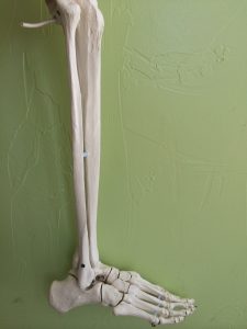
Where are the fibularis muscles?
Using the word fibularis makes more sense to me than peroneus because the three muscles that we are talking about all attach to the fibula, which is the smaller of the two bones in your lower leg. The fibula looks like the bow for a violin.
Once you learn about the fibula bone you’re set, you’ve actually learned one less word. Now we’re talkin’!
All three fibularis muscles attach to the fibula on one side and to the foot in different places on the other, which is how they help the ankle move and stretch.
Find each of these muscles on yourself as we review the attachments.
Fibularis Tertius attachments – This is the smallest muscle of the three. It begins at the lowest (inferior) portion of the fibula and connects into the top of the 5th metatarsal. This makes it great at dorsiflexion and eversion of the ankle.
Fibularis Brevis attachments – This muscle attaches in the middle of the fibula and attaches to the outside (lateral) portion of the 5th metatarsal giving it more advantage toward ankle eversion and the ability to dorsiflex or plantarflex the ankle depending on starting position.
Fibularis Longus attachments – As the name states, it’s the longest, attaching to the most upper (superior) portion of the fibula. The other side wraps around under the outside (lateral) edge of the foot to the 1st metatarsal and cuneiform. This gives the longus the ability to influence eversion from a position of plantarflexion.
What do the fibularis muscles do?
Aside from the specific motions described above, these three muscles as a whole support the outside (lateral) ankle. On the contrary, when a client rolls their ankle, it’s usually one of these three muscles that get overstretched and injured.
The fibularis muscles evert, plantarflex and dorsiflex your ankle when walking, squatting, running, lunging, etc. making their mobility and stability both very important! Everything you do starts from your feet, so keep that base of support strong.
Once we know where a muscle attaches and the movements it creates, it’s fairly simple to design an exercise to strengthen that muscle.
Fibularis Tertius Exercise – Have your client dorsiflex and evert their ankle using just gravity or by attaching a resistance band around their midfoot to add additional resistance.
Fibularis Brevis Exercise – Have your client dorsiflex with more abduction than for tertius. Use a resistance band to challenge the eversion component.
Fibularis Longus Exercise – Have your client plantarflex, abduct and evert their ankle, using a resistance band or your hand (if comfortable with it) to create force.
Fibularis study tip
To remember these muscles better, find the attachment points on yourself. Evert, dorsiflex and plantarflex your ankle while placing your hand where the attachment sites are to feel the contraction of the muscles. Using a balloon or rubber band is a way to bring the muscle to the outside of the skin so you can better visualize what it doesn’t.
When you’re walking, pay attention to the role that these muscles have in every step you take.
Learning anatomy and muscle attachments enable you as a personal trainer to create effective exercises and cue proper form with your clients. Keeping bodies strong and your business healthy. It’s important to strengthen these muscles with the same emphasis you place on the pectoral group or the abdominals.

[info type=”facebook”How do you strengthen your clients ankles and feer? If you’re an NFPT trainer, join the Facebook Community Group to mingle with other trainers. If you’re not, come talk with NFPT here, we would like to meet you![/info]
References:
Abrahams, P.H. et al. 2003. McMinn’s Color Atlas of Human Anatomy. London: Elseiver.
Muscolino, Joseph E. 2004. Musculoskeletal Anatomy Coloring Book. Philadelphia, PA: Mosby, Inc.
Beverly Hosford, MA teaches anatomy and body awareness using a skeleton named Andy, balloons, play-doh, ribbons, guided visualizations, and corrective exercises. She is an instructor, author, and a business coach for fitness professionals. Learn how to help your clients sleep better with in Bev's NFPT Sleep Coach Program and dive deeper into anatomy in her NFPT Fundamentals of Anatomy Course.